The Polygraph Process
Chart Collection Phase
During the chart collection phase, the examiner administers the examination and records physiological data while the examinee responds to the relevant questions. Several sensors are attached to the examinee in order to monitor physiological activity throughout the testing process.
These may include:
- pneumograph sensors to monitor respiration
- a blood-pressure cuff to monitor cardiovascular activity
- electrodermal sensors to monitor skin conductivity changes
Data Analysis Phase
Following chart collection, the examiner analyzes the physiological data and renders a professional opinion based on the examination results and observed chart activity.
Polygraph Sensors Explained
Modern Polygraph Instrumentation
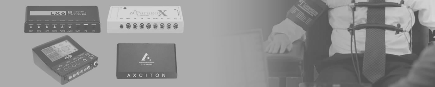
Polygraph instrumentation has evolved significantly over the past several decades. Earlier analogue systems relied on mechanical components and paper chart recordings, often depicted in popular media as pens tracing lines across a continuously moving paper roll.
Modern polygraph examinations are typically conducted using computerized digital instrumentation. Physiological data is collected electronically and displayed in real time on a computer workstation, allowing for detailed analysis using established examination methodologies and scoring procedures.
During the examination, a number of sensors are placed on the examinee in order to monitor specific physiological activities. These sensors are positioned in a non-invasive manner and are designed to record physiological data throughout the testing process.
The polygraph does not directly detect deception. Rather, it records and displays physiological responses associated with the examination. A trained examiner evaluates fluctuations and patterns within the recorded data and interprets those findings in the context of the examination procedure and the issues under consideration.
The examination process combines professional interviewing techniques, standardized testing procedures, and physiological data analysis to assist in credibility assessment and investigative decision-making.
Respiratory Rate
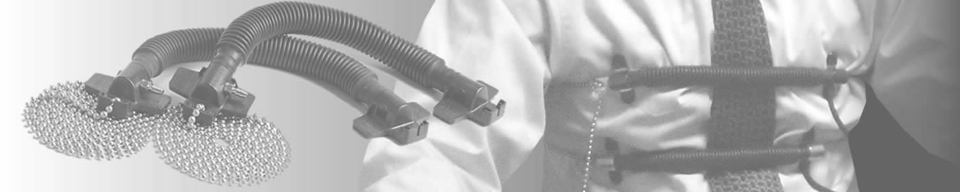
Two pneumographs, rubber tubes filled with air, are placed around the examinee’s chest and abdomen.
When the chest or abdominal muscles expand, the air inside the tubes is displaced. In an analog polygraph, the displaced air acts on a bellows, an accordion-like device that contracts when the tubes expand.
This bellows is attached to a mechanical arm, which is connected to an ink-filled pen that makes marks on the scrolling paper when the subject takes a breath.
A digital polygraph also uses pneumographs, but employs transducers to convert the energy of the displaced air into electronic signals.
Cardiovascular Activity / Heart Rate Variation
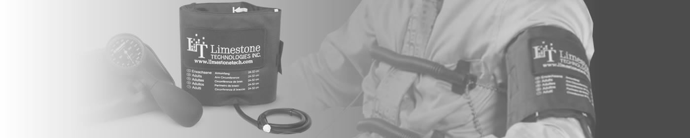
A blood-pressure cuff is placed around the subject’s upper arm. Tubing runs from the cuff to the polygraph. As blood pumps through the arm, changes in pressure displace the air in the tubes, which are connected to a bellows that moves the recording pen. In digital polygraphs, these signals are converted into electrical signals by transducers.
Electrodermal Activity
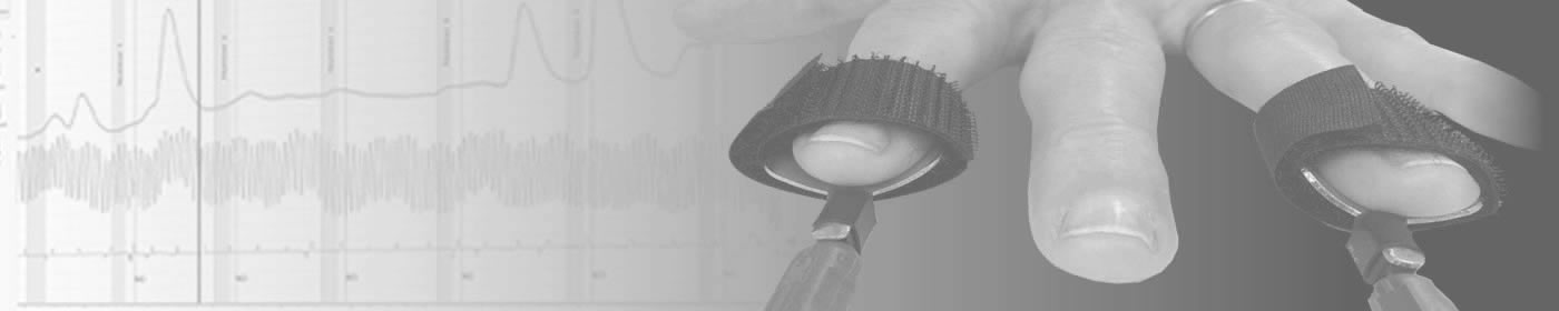
To simplify, electrodermal activity is a measure of changes in skin conductivity, often associated with moisture or sweat activity on the fingertips.
The fingertips are among the more responsive areas of the body for this type of measurement and are therefore commonly used for electrodermal monitoring.
A person may sweat more when experiencing stress, arousal, or significant cognitive activity.
Fingerplates, called electrodermal activity sensors, are attached to two of the subject’s fingers.
These plates measure the skin’s ability to conduct electricity.
When the skin is hydrated, as with sweat, it conducts electricity more easily than when it is dry.
Vasoconstriction and Vasodilation
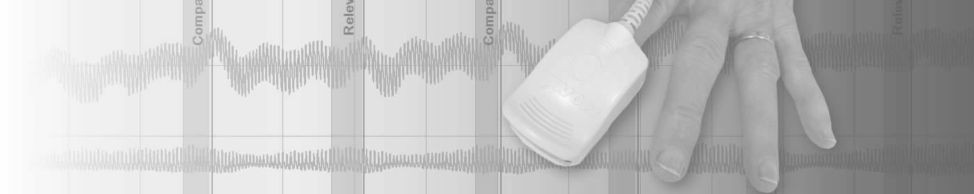
The plethysmograph sensor is typically attached to a fingertip and monitors peripheral blood flow and pulse activity throughout the examination. Using photoplethysmography technology, the sensor records changes in blood volume within the small blood vessels of the extremities and provides information relating to pulse rate, pulse amplitude, vascular activity, and blood oxygen saturation.
Changes in physiological or emotional arousal may be accompanied by vasoconstriction, the narrowing of blood vessels, or vasodilation, the widening of blood vessels.
These vascular changes can influence blood flow patterns and are reflected in the data collected by the sensor.
The plethysmograph is not evaluated in isolation. Rather, it forms part of the broader physiological data set collected during the examination and may provide additional information to assist the examiner in the analysis and interpretation of recorded physiological activity.
Polygraph Outcomes
Polygraph outcomes generally fall into the following categories:
- No Deception Indicated (NDI) / No Significant Response (NSR) - The physiological data collected during the examination was evaluated as being consistent with truthfulness regarding the issues under examination.
- Deception Indicated (DI) / Significant Response (SR) - The physiological data collected during the examination was evaluated as being consistent with deceptive responding regarding one or more issues under examination.
- Inconclusive (INC) - The recorded physiological data did not support a definitive opinion and could not be interpreted as either deceptive or non-deceptive.
- No Opinion (NO) - Due to physiological, medical, or examination-related factors, the examiner was unable to render a professional opinion regarding the examination results.
- Purposeful Non-Cooperation (PNC) - The examinee's actions were determined to have interfered with the examination process to such an extent that a valid professional opinion could not be rendered.
Conclusion
Polygraph examinations are widely used internationally in:
- criminal investigations
- law-enforcement screening
- integrity management programs
- employment screening
- commercial investigations
Polygraph examinations are intended to function as one component within a broader investigative or decision-making process rather than as a replacement for conventional investigative methods.
Questions regarding examination procedures or polygraph testing may be submitted through the .